Display Spectrometer
A special photoelectron analyzer displays an image of the photoelectrons emitted in various directions. With the high brightness at an undulator at the ALS such images take only a few seconds to acquire. That makes it possible to quickly explore electrons emitted in all directions and thereby obtain a complete picture of electrons in solids and at surfaces.

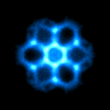
Emission pattern of photoelectrons from the (111) surface of diamond, measured in two seconds at the ALS.
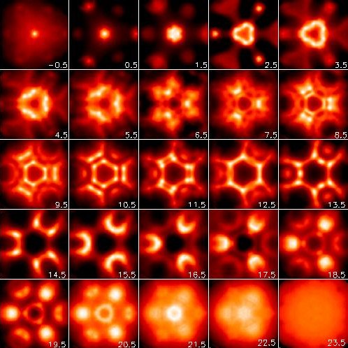
The most common method for describing electrons in solids is local density theory. The 1998 Nobel Prize in Chemistry was awarded for the development of this method. It works so well that it has been difficult to test its limits and to find ways to take its accuracy to the next level. As fundamental characteristic of electrons in solids one can choose their band width. It reflects the strength of the chemical bonds holding the solid together. This quantity can be obtained from the emission pattern of the photoelectrons at various electron and photon energies (using synchrotron radiation). The data for diamond shown below achieve 1% accuracy, ten times better than previous experiments. They show that the band width comes out 7% too small in local density theory. It is well-described by quasiparticle calculations, a more advanced description of electrons in solids. This type of measurement has been performed in the references below for three prototypical solids, an insulator (lithium fluoride), a semiconductor (diamond), and a semi-metal (graphite).
Examples:
LiF: Himpsel et al., Phys. Rev. Lett. 68, 3611 (1992);
Diamond: Jimenez et al., Phys. Rev. B 56, 7215 (1997);
Graphite: Heske et al., Phys. Rev. B 59, 4680 (1999).

Images of photoelectrons from diamond at various energies below the top of the valence band (in eV).
The Valence Electrons in Solids and Molecules
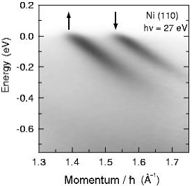
The valence electrons are the most energetic electrons in solids and molecules. They determine many important properties, such as electrical conductivity, superconductivity, magnetism, magnetoresistance, and chemical reactivity. Their energy allows them to move around in solids and to initiate chemical reactions in molecules. In solids, only a narrow energy slice is relevant. Its width corresponds to the thermal energy, which is about 0.1 eV at room temperature. The energy resolution of photoelectron spectrometers has improved such that electrons can now be probed at low temperatures with milli-eV resolution. Angle-resolved photoemission provides not only their energy, but also their momentum k. A plot of energy versus momentum ("energy band dispersion") characterizes valence electrons in solids.

Energy band dispersion of the most energetic electrons in nickel, measured with high resolution at the Synchrotron Radiation Center (SRC) in Madison. The magnetic splitting between spin up and spin down electrons is clearly resolved. The slope of the dark energy-versus-momentum lines corresponds to the electron velocity. From Altmann et al., Phys. Rev. Lett. 87, 137201 (2001).
X-ray Absorption and Emission Spectrosopy
Synchrotron light sources provide tunable X-rays and thereby enable X-ray absorption spectroscopy (XAS). This technique measures excitations from the core level of a specific atom into its empty valence orbitals. As the resulting core hole gets re-filled, the atom emits either electrons or X-rays. Electrons dominate for shallow core levels, which are the sharpest. The characterisctic fluorescence photons emitted by deeper (but broader) core levels are needed for detecting dilute atoms, such as in catalysts or for environmental contaminants. XAS of the sharpest core levels is difficult with fluorescence detection because of the small cross section, but it has become possible using undulators. The spectral range of the ALS matches the sharpest core levels of semiconductors, organic materials, biomolecules, and magnetic materials (see a table of the sharpest core levels of the elements and their binding energies).
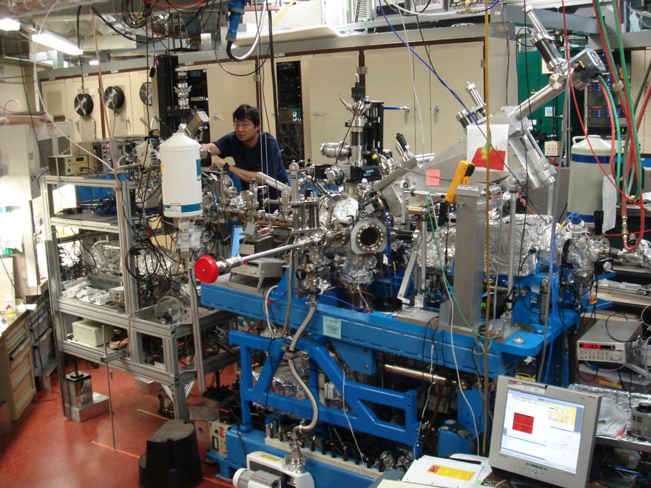
XAS has been used for a wide variety of applications. A few examples are given in the references below. The image shows the downstream part of a beamline at the ALS with two endstations in series. A bendable upstream mirror allows a wide range of focal points located at various endstations. For radiation-sensitive organic and bio-materials the light can be defocused as well. Various photon detectors enable an optimized trade-off between spectral selectivity and detection efficiency.
Examples:
Buried layer of B dopants in silicon:
Carlisle et al., Appl. Phys. Lett. 67, 34 (1995)
Magnetic nanocrystals protected by organic molecules:
Perez-Dieste et al., Appl. Phys. Lett. 83, 5053 (2003)
Orientation of DNA molecules attached to a surface:
Petrovykh et al., J. Am. Chem. Soc. 128, 2 (2006)
Protein immobilized at a surface:
Liu et al., Langmuir 22, 7719 (2006)
Dyes for biomimetic solar cells:
Cook et al., J. Chem. Phys. 131, 194701 (2009).
Atom-specific orbitals in a donor-absorber-acceptor complex:
Zegkinoglou et al., J. Phys. Chem. C 117, 13357 (2013).

Two endstations in a row for photon in / photon out spectroscopy at the ALS.
Supported by NSF-DMR, NSF-CHE, and DOE-BES.
Franz Himpsel's Home Page